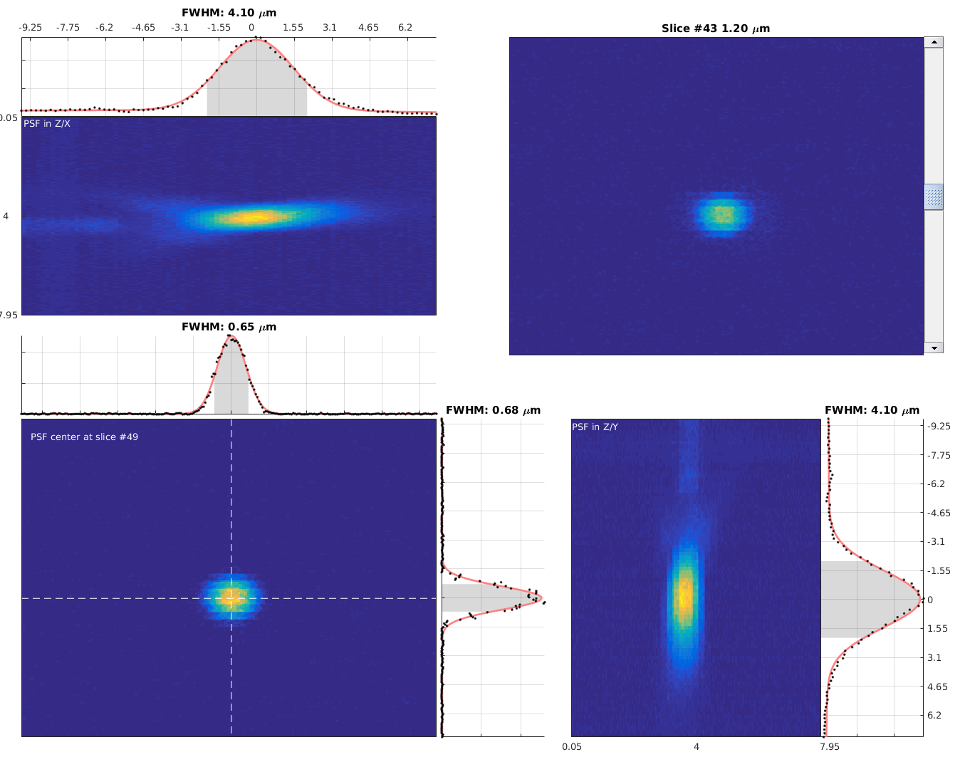
This repository contains various tools for measuring microscope performance with ScanImage. In particular, there are functions for recording and measuring Point Spread Functions (PSFs), and for measuring field of view size using an EM grid.
mpqc.record.PSFto easily acquire PSF stacks with ScanImage.mpqc.record.lens_tissueto acquire standardised data from lens tissue.mpqc.record.standard_light_sourceto data from a standard light sourcemeasurePSFto estimate PSF size. For a demo, runmeasurePSF('demo').Grid2MicsPerPixelmeasures the number of microns per pixel along x and y by analyzing an image of an EM grid.mpqc.tools.meanFrameplots the mean frame intensity as a function of time whilst you are scanning.
Add the multiphoton-qc code directory to your MATLAB path.
You do not need to "Add With Subfolders".
Output files contain meta-data associated with the microscope in order to make it easier to compare different microscopes and to track hardware changes.
Before using the tools for the first time you must run mpqc.settings.readSettings; and then you must fill in the YAML file for your PC.
The commands which record information assume this file has been created and filled in with your microscope details.
Future changes to the software may add fields to this file automatically.
If this happens, it will be reported to the CLI that a new field was added with default settings.
If you don't change the defaults, probably nothing bad will happen but the information provided by that field will not be logged.
To get a good image of a sub-micron bead:
- Use image sizes of about 256x256 to 512x512 pixels.
- Once a bead is found, zoom to about 20x or 25x.
- Take a z-plane every 0.25 microns and average about 25 to 40 images per per plane. It's not worth doing more because we'll be fitting a curve to z-intensity profile.
To measure the PSF you can either use the included mpqc.record.PSF function or manually set up ScanImage (see documentation folder if you need to do that).
- Find a bead, zoom in.
- Select only one channel to save in the CHANNELS window.
- Set the averaging to the quantity you wish to use in the Image Controls window.
- Move the focus down to the lowest point you wish to image and press press "Zero Z" in MOTOR CONTROLS
- Now focus back up to where you want to start the stack and press "Read Pos" in MOTOR CONTROLS. This is the number of microns you will acquire (ignore the negative sign if present).
- Run
mpqc.record.PSFwith number of microns obtained above as the first input argument. This will obtain the stack with a 0.25 micron resolution using the averaging you have set. e.g.record.PSF(12)for a 12 micron stack. The save location is reported to screen. You can define a different z resolution using the second input argument.
To view the PSF run measurePSF and load the saved stack using the GUI. For more info type help measurePSF.
If loading ScanImage TIFFs the voxel size is extracted automatically from the header information in the file.
Otherwise, this information can be provided using the second and third input arguments (see help measurePSF).
See documentation folder for how to make an EM grid slide.
To image the slide:
- Copper will autofluoresce when illuminated by a 2-photon laser.
- Use any 2p wavelength and very low power e.g. 3 mW.
- The grid should be oriented so that it's aligned relatively closely with the scan axes (i.e. the edges of the image).
This will make it easier to see distortions by eye and also to run
Grid2MicsPerPixel. - Hit "Focus" in ScanImage. Acquire data with just one channel. Get a good clear grid image and average a few frames if needed.
- Press "Abort" to stop scanning once a good image is obtained.
- Run
Grid2MicsPerPixelto measure the FOV. The function will automatically pull data from ScanImage. Look at the diagnostic figures to ensure function has found most of the grid lines (it uses the median grid line distance so you don't need all the lines). If very few grid lines are detected, the results will be meaningless. - You can use the buttons in the GUI to obtain new images.
- Once you are happy with the results you can use the "Apply FOV" button to calibrate ScanImage.
Run mpqc.tools.meanFrame to bring up a figure window that plots mean frame intensity during scanning.
This function is used for things like tweaking a pre-chirper.
See help mpqc.tools.meanFrame for advanced usage.
To record data:
mpqc.record.uniform_slideThe results are saved to a folder on the desktop. You can view the results as follows:
% cd to directory containing data
mpqc.plot.uniform_slide('uniform_slice_zoom_1_920nm_5mW__2022-08-02_10-09-33_00001.tif')Remove all contaminant sources of light from the enclosure run:
mpqc.record.electrical_noiseLens paper at a known laser power is a reasonable choice for a standard structured target. If laser power is carefully checked before the recording, it can be used to compare different rigs. This is because we can convert the data to photons. If a standard source is imaged after the lens paper at the same gains, it can be converted to photons to compare the detection paths of different rigs. NOTE: these functions work but they are not finalised and documentation is sparse right now.
First record the lens paper. Do this at around 50 to 100 mW at the sample. The following command will record the lens paper at 4 different gains. The gains are set automatically AND HAVE ONLY BEEN TESTED WITH MULTI-ALKALI PMTs SO FAR.
mpqc.record.lens_paperThen take out the lens paper. CLOSE THE LASER SHUTTER. Record the standard source. This by default will record at the same gains as above.
mpqc.record.standard_light_sourceAnalyse this:
OUT = mpqc.analyse.get_quantalsize_from_fileThis will analyse one file at a time: returning the standard source mean photon count.
The quality of the fit that underlies this should be assessed with mpqc.analyse.plotPhotonFit.
You can generate a PDF report of all conducted analyses using
>> generateMPQCreportThese functions have been tested on most MATLAB releases R2016b and later. They should also work on R2016a but are known to fail on 2015b and earlier.
Requires the Curve-Fitting Toolbox, the Image Processing Toolbox, and the Stats Toolbox. The MATLAB Report Generator is needed if you want to make PDF reports. It is known to work with ScanImage Basic 2020 to 2022.
Please see the list of known obvious issues before using the software.
